


Varicose veins on the lower part of the legs are characterized by the expansion of the superficial veins of the legs, which are accompanied by a violation of blood flow in them and valve failure. As a result, the veins increase in length and diameter, acquiring the appearance of a serpentine, cylindrical or sacular, although there are also mixed manifestations of the listed defects.
Characteristics of the venous system
The emergence and development of varicose veins is directly related to the venous system of the legs, which consists of:
- saphenous vein: small and large;
- veins located in (in the lower part of the legs and thighs);
- perforated vein, which is the link between the two previous systems.
Typically, 90% of blood is sent to the bottom of the leg through deep veins, and the remaining 10% through superficial surfaces. Upon returning to the heart, this mechanism is supported by valves in the venous wall. When the next part of the blood arrives, they slam to prevent its movement from top to bottom under the influence of gravity. Muscle contraction pushes blood further into the heart, allowing normal blood flow.
With long -term a person in an upright position, blood stasis can develop, which increases the pressure on the vein and causes an increase in its diameter. This process provokes incomplete closure of the leaflets of the valve, as a result of which blood flow is disrupted by the return flow from the heart - reflux.
The venous valve is most likely affected, as it transports the most amount of blood and therefore experiences maximum load. To lower the high pressure in it, a portion of the blood is transported by perforated veins to a superficial surface, which is not initially intended for large volumes. Such a load on the venous wall leads to the expansion and formation of varicose veins.
At the same time, blood enters the deep veins without stopping, but due to violation of its function and the normal activity of the valve leaves from the perforated veins, the blood is distributed to the superficial channels. As a result, chronic varicose veins develop, which over time are accompanied by painful sensations, edema and trophic ulcers.
The cause of the disease
Previously, one of the main causes of varicose veins was called hereditary factors, but today this theory has been disputed. Of course, it is possible to trace the manifestations of the disease that often occur in some families, but this is more likely due to the peculiarities of life inherited in the family: food culture, passive rest, inactive work, and the like.
The development of varicose veins is based on the presence of reflux in the venous system, when blood circulates through the veins in the opposite direction. Additional blood transport from distant veins to superficial veins is possible due to congenital degenerative pathology or acquired valve apparatus. This causes too many superficial blood vessels with blood and their distance when venous nodes form.
One of the basic reasons for the development of varicose veins is considered to be an unhealthy diet, which in some cases leads to obesity. People like this move a little, especially eating processed foods, and the portion of plant fiber in the food is minimized. However, they are the ones involved in strengthening the walls of veins and blood vessels and preventing prolonged chronic constipation, which greatly increases intra-abdominal pressure and thus provokes varicose veins. It has been observed that weight gain of more than 20% increases the risk of disease fivefold.
The main provoking factor for women is childbirth, while the risk of varicose veins increases with each subsequent pregnancy. Severe weight gain and an enlarged uterus put a lot of pressure on the legs, which are stagnant. This condition is exacerbated by the ever-increasing intra-abdominal pressure and the action of the hormone progesterone, which affects the state of the elastic fibers in the walls of blood vessels.
Other factors that provoke varicose veins in the lower extremities include:
- a sedentary lifestyle, standing upright during the day (e. g. , hairdressing), long flights or long trips. All this leads to a stagnant process in the lower part of the leg, when blood accumulates in the superficial veins and is less carried to the heart;
- sometimes increases the risk of developing varicose veins for women, wearing uncomfortable and tight shoes, especially models with high heels;
- tight corsets and panties squeeze the inguinal veins and increase intra-abdominal pressure, which is a direct prerequisite for varicose veins;
- high blood pressure;
- smoking, which indirectly leads to thinning of the walls of blood vessels.
Classification of diseases
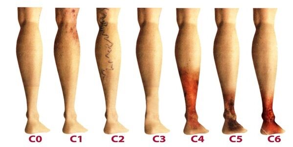
Varicose veins in the lower leg are classified depending on the prevalence of venous lesions, their localization, as well as the presence of pathological reflux, which is characterized by disturbances of blood flow. There are 4 forms of varicose veins:
- intracutaneous and subcutaneous (segmental) varicose veins, in which there is no pathological venous blood outflow;
- segmental varicose veins, when reflux occurs through perforated or superficial veins;
- a common form of varicose veins, in which reflux occurs through perforated and superficial veins at the same time;
- varicose veins are characterized by reflux in the deep veins.
Once varicose veins in the lower legs become chronic, phlebology considers three degrees:
- Temporary edema, occurring periodically against the background of "heavy foot" syndrome.
- Persistent, persistent edema. Hyperpigmentation and eczema may appear.
- Trophic venous ulcers.
The latter stage is the most difficult to treat, as it requires early removal of inflammation and healing of skin tissue.
Stages and symptoms
The disease develops very slowly, sometimes over a dozen years, so that the symptoms that appear will force the patient to seek advice from a phlebologist. In the early stages of varicose veins, manifestations are often associated with fatigue, age, or other causes. To fully consider the symptoms of the disease, the manifestations are classified according to the stage of varicose veins:
- The first stage begins to manifest itself more often at a young age - after 20 years, when there is a feeling of heaviness in the legs, edema may appear, which disappears completely overnight. At the bottom of the lower leg, you can see enlarged veins, indicated by a thick protrusion of skin. At this stage, many people see small spider veins. In general, symptomatic are subtle and rarely receive due attention.
- The second stage is characterized by increased external manifestations of dilated veins. The disease has already developed against the background of pathological work of the venous valve, therefore, the saphenous vein increases significantly, and its elongation can also be observed. More often there is a feeling of heaviness and burning in the feet, they quickly get tired of walking.
- The disease is already chronic due to a constant imbalance in venous blood flow. At night, patients experience edema of the near ankle, which can be very intense. There is a feeling of heaviness in the legs, and cramps may occur at night.
- If there is no treatment at an earlier stage, chronic insufficiency of venous system function has a negative impact on metabolic processes in the skin, the area in the lower part of the foot is severely affected. Darkening of the skin appears near the ankles - hyperpigmentation, it thickens and becomes inflamed over time. The condition described is called lipodermatosclerosis. If at this time you do not start therapy with respect to the venous system, then trophic ulcers will immediately form.
- The fifth stage is accompanied by many trophic ulcers, some of which heal periodically with scar formation.
- In zones of trophic disorders that have long existed, extensive ulcers open. This condition requires immediate active therapy, aimed at the treatment of varicose veins and the healing of skin ulcers.
Diagnostics
External examination of the lower extremities in the vertical and horizontal positions of the body, palpation of the veins and initial assessment of the stage of the disease were performed. The patient is sent for a general blood test, which allows you to study the picture of the disease more closely:
- at the platelet level, the predisposition to thrombosis will be reflected;
- hemoglobin levels, as well as red blood cell counts, indicate blood clotting levels;
- with increased levels of leukocytes, one can assess about inflammation, which helps diagnose thrombophlebitis more quickly.
Be sure to check the venous system of the legs, for which there are many methods:
- ultrasound dopplerography - USDG;
- phlebography;
- CT phlebography;
- duplex angioscanning - USAS;
- phleboscintiography;
- photoplesmography;
- phlebomanometry and the like.
In practice, patients are more often prescribed USAS and ultrasound, as they help fully study the leg venous system and identify degenerative areas. The remaining methods can be prescribed in addition if the ultrasound examination does not give a complete picture of the picture of the disease. Some of these methods can cause complications such as venous thrombosis, perforation of the vessel wall with a catheter, and allergy to contrast agents. Consider the most commonly used techniques in phlebology:
- USAS allows assessing the anatomical, hemodynamic and functional pathology of the venous bed. The data obtained are subject to computer processing, after which a model of the venous system can be viewed on video or printed on paper.
- Doppler ultrasonography with high accuracy determines the patency of superficial veins and is located in, the velocity of blood flow. Doppler ultrasonography makes it possible to assess the function of the valve device.
After performing an extensive diagnostic, the doctor creates a patient’s phlebocard, which allows you to determine the segment of the damaged venous system, its extent and length. After that, the appropriate treatment is selected.
Treatment
It is done comprehensively and determined based on symptoms, stage of disease progression and study results. In the early stages, conservative therapy is prescribed, which consists of:
- Drug treatment when a group of drugs is prescribed:
- antioprotectors and phlebotonics;
- anticoagulant;
- divisor
- topical preparations (ointments, gels);
- anti-inflammatory drugs.
- Elastic compression, used for wearing socks or compression bandages (rare). It allows you to squeeze the muscles, prevent the process of stagnation, increase blood flow through the ducts. When wearing such underwear, there is the effect of artificially maintaining the vascular tone.
- Physiotherapeutic methods, among which the best treatment results are shown by electrophoresis, diadynamic currents, laser radiation and magnetic fields.
- Appropriate physical activity, which should be done only with clothing in compression (except for swimming). Cycling, swimming, jogging are recommended. The phlebologist selects a set of exercises for the lower leg, which will train the foot channel on a daily basis.
In addition, patients are advised to perform a different five -minute bathing procedure each night, alternately switching from warm water to cold water. Such manipulation improves blood flow and tone of blood vessels.
It is important at the beginning of treatment to identify the factors that provoke the disease in order to influence it effectively. And patients at risk should visit a phlebologist every 2 years for a preventive examination and perform an ultrasound examination of the veins in the legs.
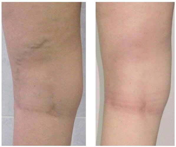
When conservative treatment does not give results or varicose veins are observed at an advanced stage, then surgical intervention is used. Today varicose veins can be completely cured by the following methods:
- Phlebectomy. The essence of the operation is to remove the main trunk of the superficial vein to eliminate pathological bleeding. Perforated veins are often entangled for the same purpose.
- Sclerotherapy. This consists of the introduction of sclerosant into the affected vein area, which leads to its wall connection. Recently, they began to actively use foamed sclerosant for the same purpose according to the technology -. Blood flow through the damaged area stops and cosmetic defects in the form of prominent nodules are removed. After such an intervention, no scars were left, all manipulations were performed on an outpatient basis without inpatient treatment. But sclerotherapy is only used for the consolidation of small branches of the venous trunk.
- Laser freezing. With the help of a laser beam, the marked part of the vein is heated, its walls stick together and the blood flow through it stops. But this technique is indicated only for veins with an expansion diameter of less than one centimeter.
prevention
Preventive measures can be primary, aimed at preventing the development of varicose veins, and secondary, when necessary to reduce the risk of relapse after surgery or to prevent worsening of the disease. Helpful hints:
- lead an active lifestyle without heavy loads on the legs: swimming, walking, cycling;
- keep an eye on your weight;
- make sure both legs are lifted more often;
- do not wear tight underwear and heels exceeding 4 centimeters;
- use orthopedic soles;
- bath with contrast;
- do preventive leg exercises for five minutes every day;
- wear compression stockings for long walks.
If you notice the slightest suspicion of varicose veins - prominent nodules on the legs, swelling, heaviness, then do not delay a visit to a phlebologist. Indeed, over time, this dangerous disease can lead to many complications, including thrombophlebitis and thrombosis.